

doximityask
May 13, 2026
Turn a Wound Photo Into a Differential

# Dermatology
# Ekgs
# Cardiology
# Uploads
# Attachments
# Xray
Clinical photos, EKGs, and imaging become much more useful when paired with AI, patient history and the right clinical question.

Doximity Team
Doximity Ask can describe and interpret visual content including EKGs, X-rays, wound photos, rash photos, and echo report screenshots. How useful that interpretation is depends entirely on the context you provide.
The pattern that works best for image uploads mirrors what you would tell a consulting colleague: who the patient is, what you are looking at, and what clinical question you need answered. Vague prompts produce vague output. Specific prompts produce specific, actionable interpretations.
IMPORTANT Image interpretation in Doximity Ask is a clinical aid, not a diagnostic read. For any finding that will change management, verify with formal radiology or cardiology interpretation. |
EKG Uploads
Give Doximity Ask the patient's age, symptoms, relevant history, and current medications, especially antiarrhythmics, rate control agents, or QT-prolonging drugs. If there is a prior EKG for comparison, note any known baseline changes.
Copy this prompt: EKG interpretation: This is a 12-lead EKG from a [age]-year-old with [symptoms]. Relevant history: [PMH, cardiac meds]. Describe the rhythm, rate, axis, intervals, and any notable findings. Flag anything that needs urgent action. |
X-Ray and Imaging Uploads
Orient Doximity Ask to the clinical question. "Is this fracture displaced?" gets a more useful answer than "what is this?" You can also upload screenshots of radiology reports if you want Doximity Ask to summarize findings or explain clinical implications for a specific patient context.
Copy this prompt: X-ray assessment: This is a [body part] X-ray from a [age]-year-old who [mechanism / presenting complaint]. I'm looking to assess whether [specific finding]. Describe what you see. |
Clinical Photos: Wounds, Rashes, and Exam Findings
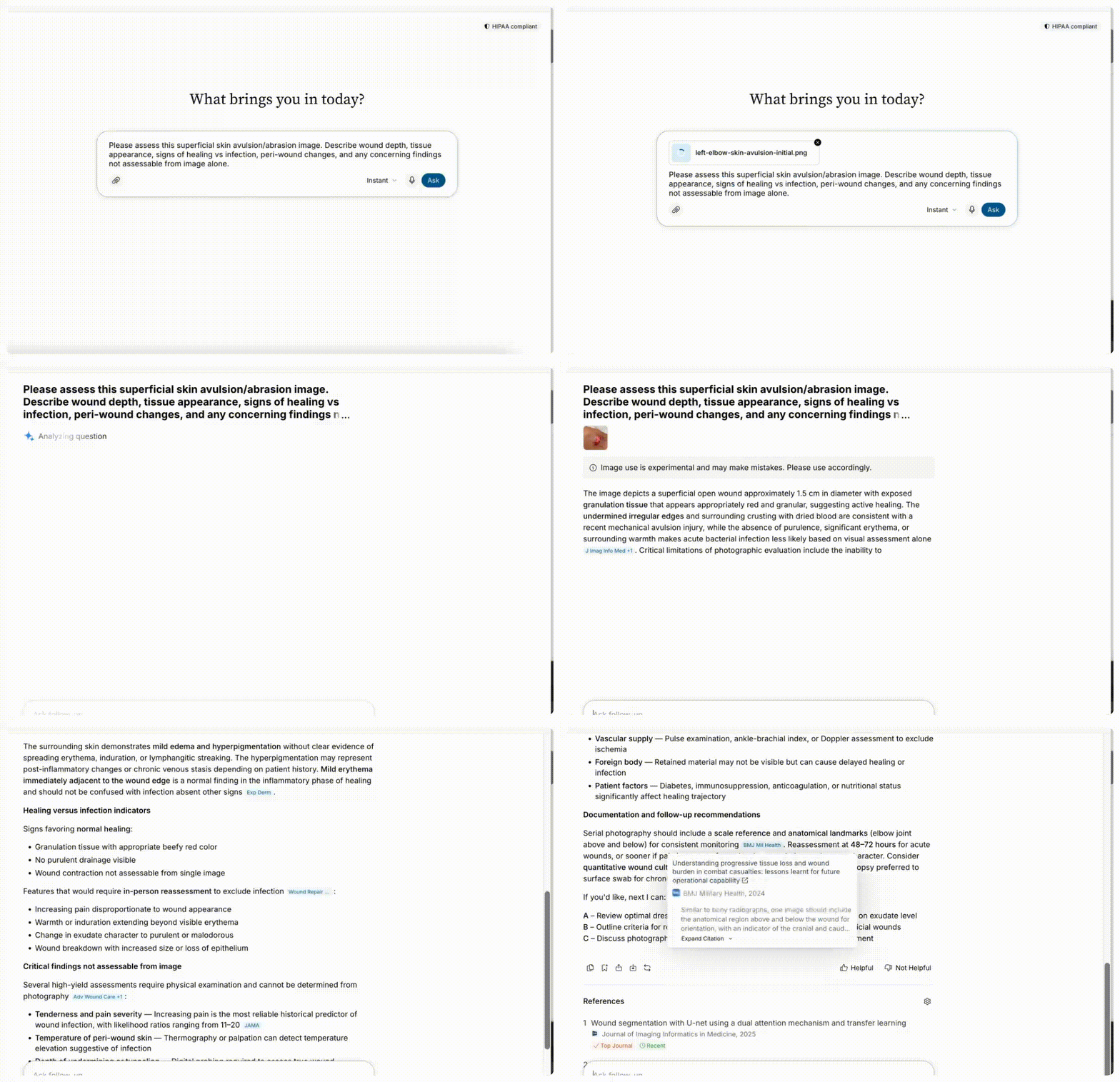
For clinical photos, describe how long the finding has been present and any relevant exposure history. Clinicians have used Doximity Ask for bedside wound photos, rashes photographed during the exam, and foot findings in diabetic patients.
Copy this prompt and upload a photo of a wound Assess this superficial skin avulsion/abrasion image. Describe wound depth, tissue appearance, signs of healing vs infection, peri-wound changes, and any concerning findings not assessable from image alone. |
Copy this prompt: Physical exam photo to documentation language: I'm uploading a photo of my patient's [body part]. Give me a description I can use in my physical exam documentation, specifically the appearance, distribution, and any features suggesting vascular, neuropathic, infectious, or other etiology. |
Dive in
Related
Education
Your Scribe Note Just Finished. Here's how to get it into your EHR.
By Doximity Team • Sep 3rd, 2026 • Views 121
Education
Turn a Dialer Phone Call Into a Scribe Chart Note, Automatically
By Doximity Team • Sep 3rd, 2026 • Views 45
Education
Turn a Dialer Phone Call Into a Scribe Chart Note, Automatically
By Doximity Team • Sep 3rd, 2026 • Views 45
Education
Your Scribe Note Just Finished. Here's how to get it into your EHR.
By Doximity Team • Sep 3rd, 2026 • Views 121